
Before and After Photos
The following are before and after photos of real Clarus Dermatology patients.
Surgical Procedures
Before
After
Keloid Tumor Excision
This gentleman was referred to our clinic for a keloid growth that had worsened after prior plastic surgery excision.
Before
After
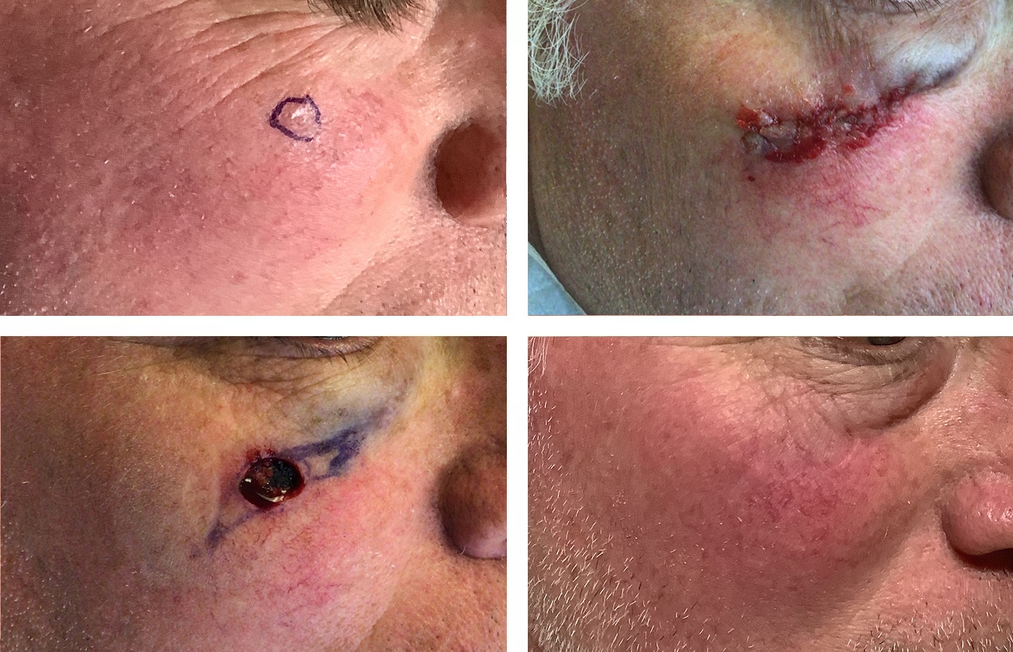
Micrographic Surgery
57 year old male with squamous cell carcinoma. Location: Left Lateral Canthus.
Before
After
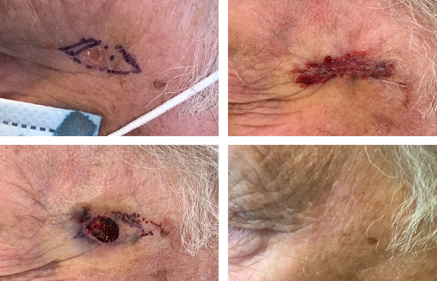
Micrographic Surgery 2
73 year old male with squamous cell carcinoma in situ. Mohs micrographic surgery. Location: Left Lateral Canthus.
Before
After
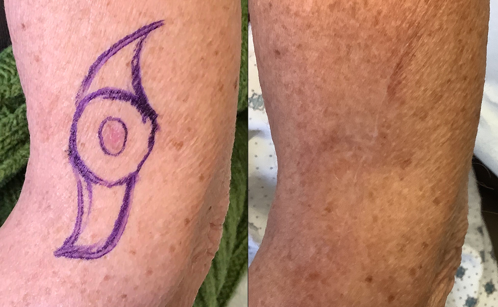
Re-Excision Left Distal Arm
66 year old female. Preoperative diagnosis: Melanoma T1A 0.4mm. Location: Left Upper Distal Arm.
Before
After
Mohs Surgery - Cheek SCC
This patient was treated for a squamous cell carcinoma in situ of the cheek. The surgical defect was closed with a linear repair.
Before
After
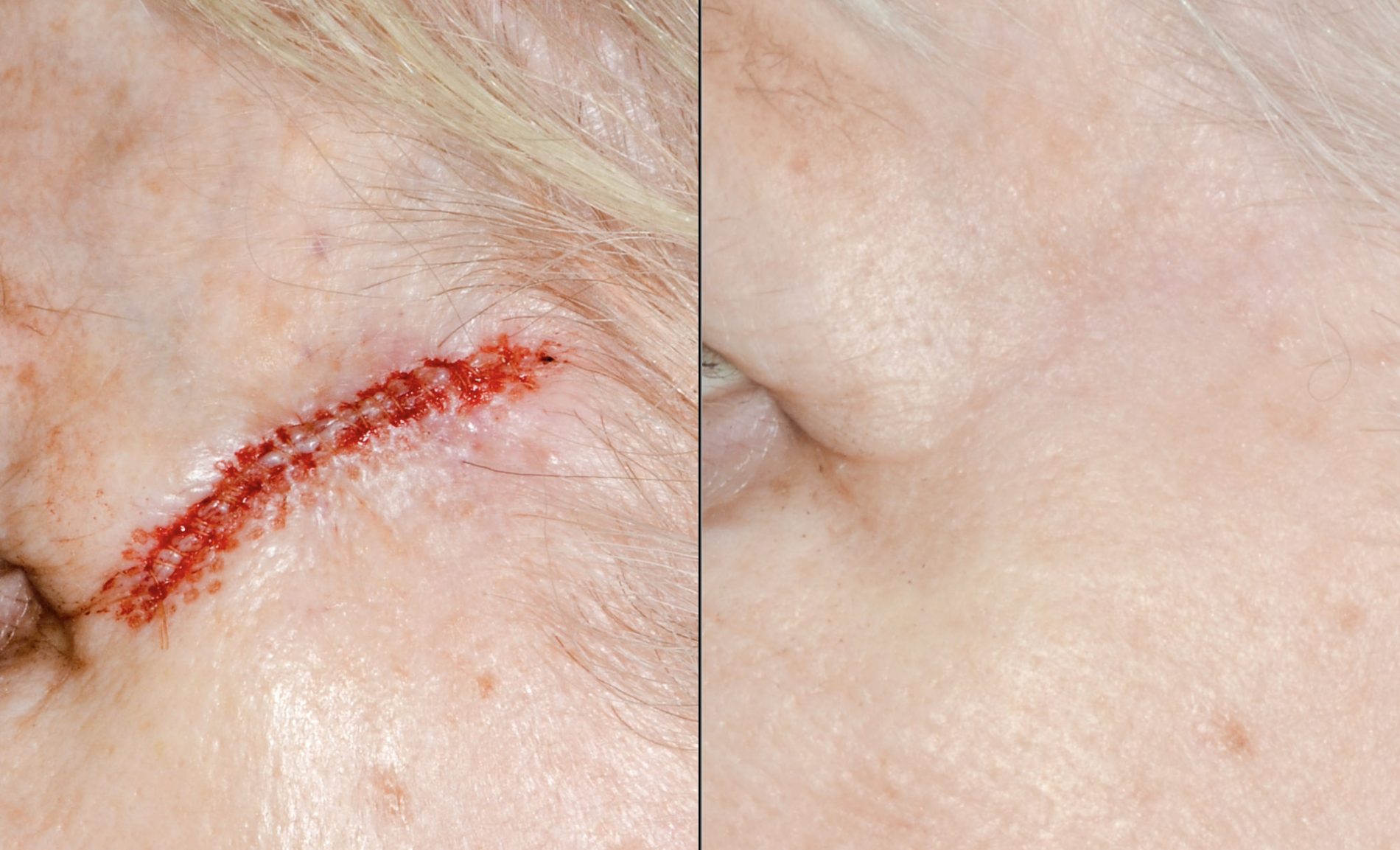
Mohs Surgery - Temple SCC
This patient was treated for a squamous cell carcinoma of the temple. The surgical defect was closed with a linear repair.
Before
After
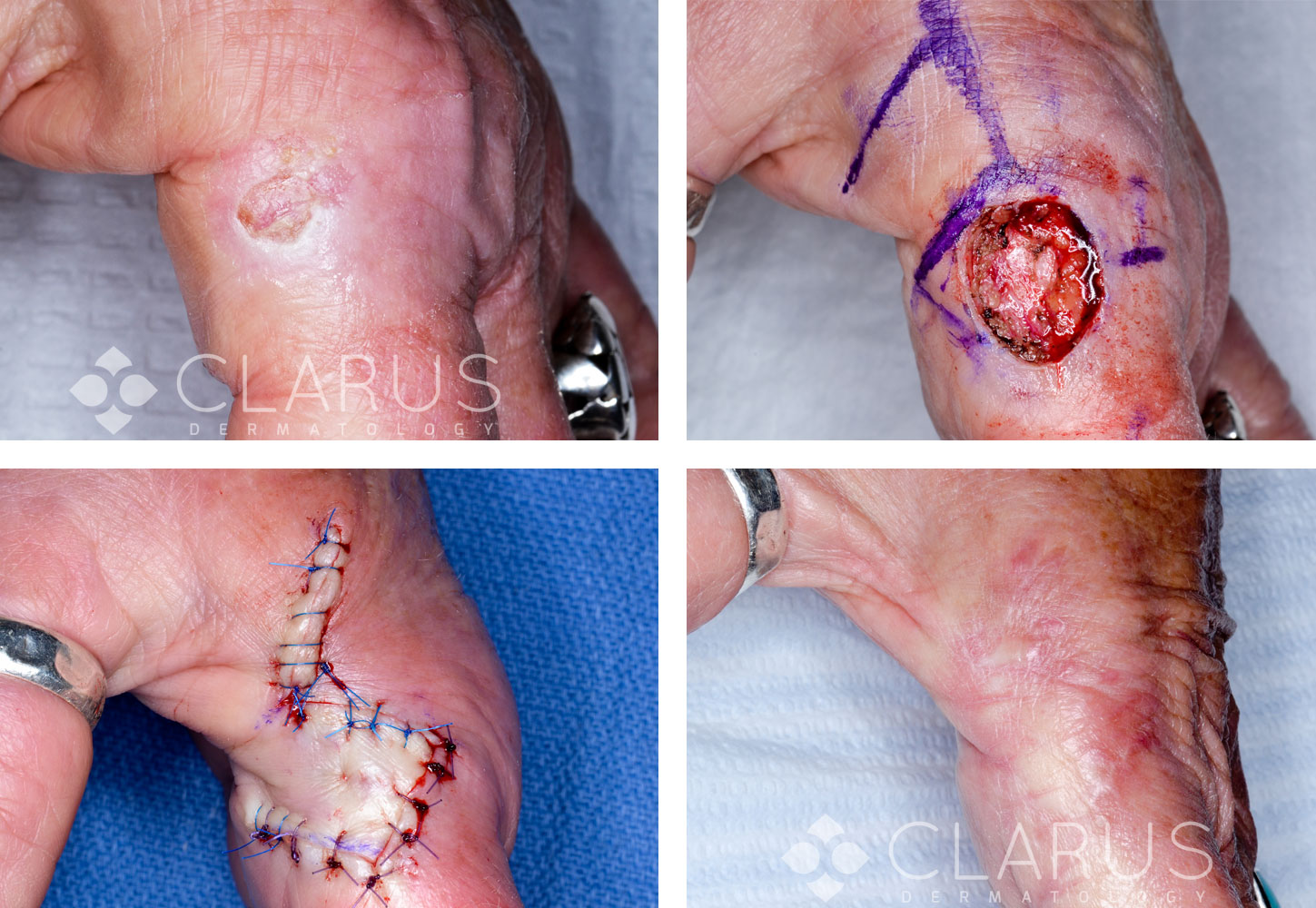
Mohs Surgery - Finger SCC
This patient was treated for a squamous cell carcinoma of the finger with Mohs surgery. A rhombic flap was used to repair the surgical defect.
Before
After
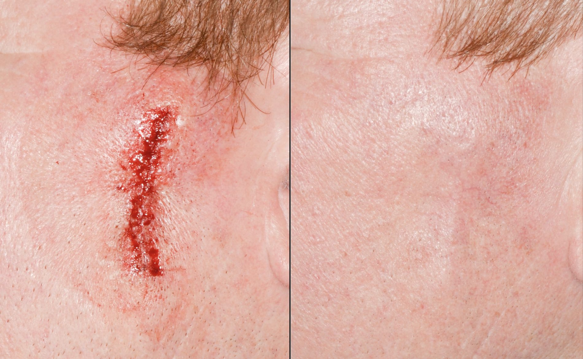
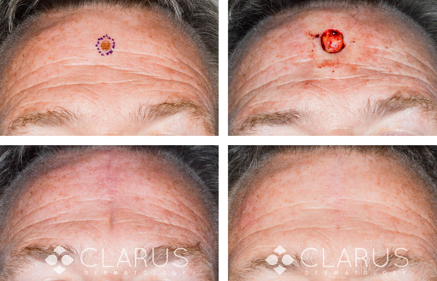
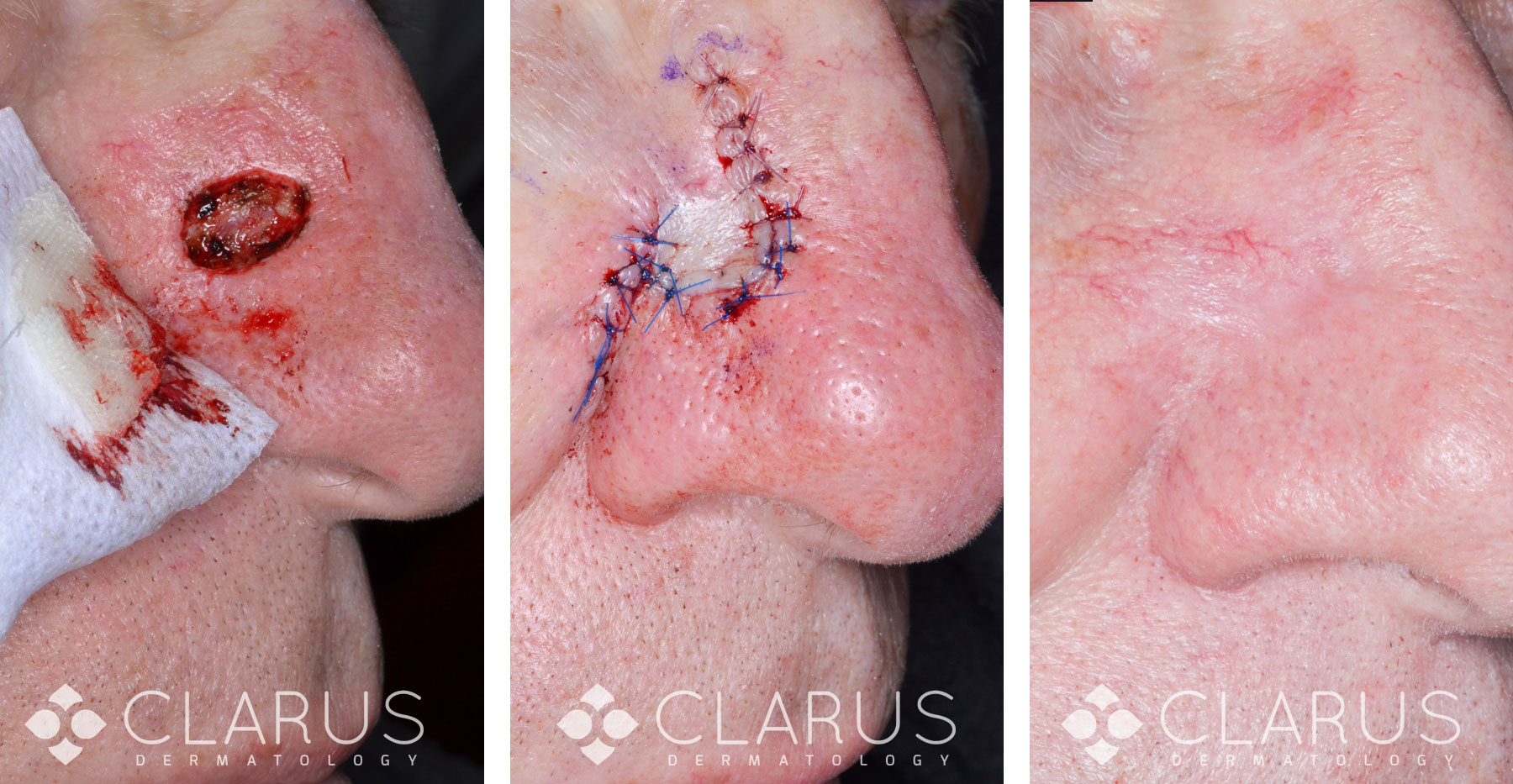
Mohs Surgery - Forehead BCC
This patient was treated for a basal cell carcinoma of the forehead with Mohs surgery. Primary closure leaves minimal visible scarring.
Before
After
Mohs Surgery - BCC Flap Reconstruction
After clearance of a basal cell carcinoma with Mohs surgery the resulting defect was reconstructed using a flap.
Before
After
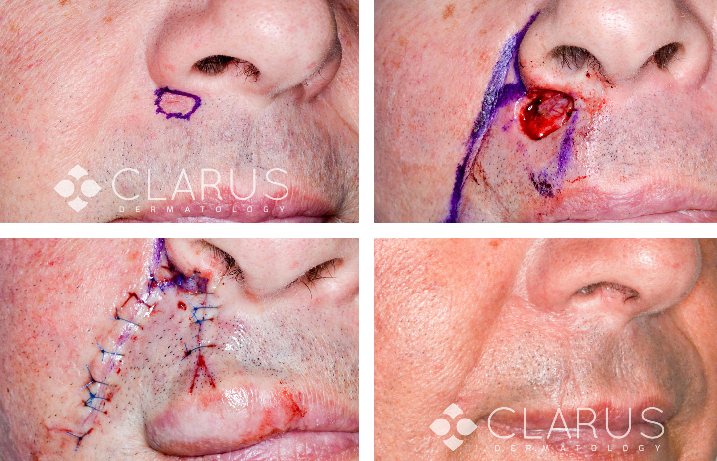
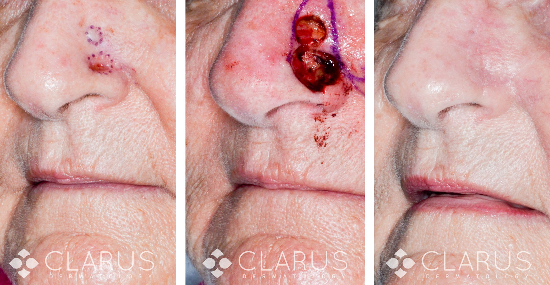
Mohs Surgery - Nose BCC
Our patient had two separate but closely spaced basal cell carcinomas on her nose. The tumors were cleared with Mohs surgery.
